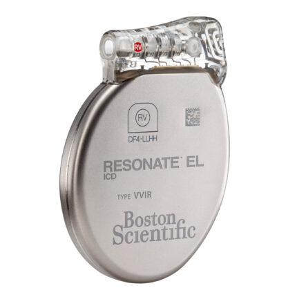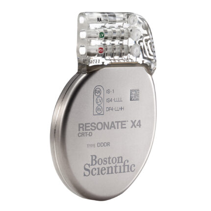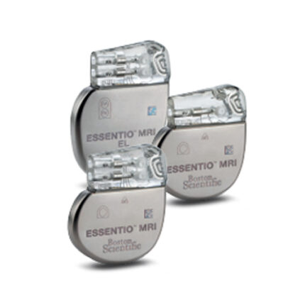

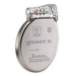
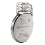

IMPLANTED CARDIOVENTRICULAR DEFIBRILLATOR TWO-CHAMBER
The MRI-compatible two-chamber implantable cardioverter-defibrillator allows patients with an implanted system to undergo 1.5T and 3T MRI scans without restrictions on the scanning area (including the heart area) or the duration of the MRI scan procedure, provided that the device is implanted with MRI-compatible electrodes and the manufacturer’s requirements for MRI scans are met.
Device: connectors: IS-1, DF-4; weight: 71.4 g.; volume: 31 cm3; Dimensions: 77 mm x 54 mm x 9.9 mm;
Materials in contact with human body tissues: Titanium, polyurethane, silicone rubber
Body shape: Physiological, thin-profile, less than 1 cm thick.
High-capacity battery with ENDURALIFE™ technology, providing a service life of more than 13 years.
The useful battery capacity of the device is 1.8 Ah.
The estimated service life is 15.4 years under the following conditions: DDDR stimulation mode, 15% stimulation, base frequency of 60 per minute, atrial and ventricular impulse duration of 0.4 ms, electrode impedance of 700 Ohm, PP/PV stimulus amplitude of 2.5 V; accelerometer on; two capacitor charges to maximum energy per year; 3-channel VPEG recording with Onset constantly on.
Wireless telemetry and wireless ECG function.
Availability of functions: automatic measurement of stimulation thresholds and automatic change of stimulation parameters when thresholds change in all
chambers.
The function of frequency adaptation.
The presence of two frequency adaptation sensors: an accelerometer and a physiological sensor for minute ventilation/respiratory sensor.
Function for adapting the AV interval to the heart rate.
Response function for AF/PT on the ventricles (ATR).
Atrial fibrillation response function (AFR).
The function of stabilizing the frequency of ventricular contractions.
Function for smoothing the rhythm frequency up and down, independently programmable.
Algorithm for reducing the percentage of unnecessary right ventricular stimulation AV Search + with search for native AV conduction.
An algorithm for reducing the percentage of unnecessary RYTHMIQ right ventricular stimulation by switching AAI(R)oCDD(R) modes (AAI(R) stimulation mode
with VVI Backup).
An advanced set of algorithms for diagnosing and monitoring heart failure: Heart Failure Sensor Suite.
AP Scan night apnea diagnostic and monitoring function.
Heart rate variability analysis function (SDANN and HRV Footprint).
The maximum programmed shock energy is 41 J.
The maximum delivered shock energy is 35 J.
The maximum accumulated shock energy is 41 J.
The standard charging time for the capacitor to reach its maximum energy (41 J) at the beginning of service is 8.4 seconds.
The maximum number of shocks per episode is 8.
Режимы стимуляции: AAI(R) with VVI Backup; DDD(R); DDD; FREE(R); NULL; VDD (R); VDD; AAI(R); AAI; VVI(R); VVI; DOO; AOO; VOO; OFF.
Parameters of stimulation. Stimulation amplitude of the PP and PG: 0.1 – 7.5 V.
Pulse width: 0.1-2.0 ms.
Automatic amplitude measurement and adjustment of sensitivity thresholds for PP and PJ.
Sensitivity of the PP and PJ: Auto (AGC), 0.15-1.5 mV.
The polarity of the pancreatic stimulation is integrated bipolar.
The rhythm hysteresis function.
The maximum transmission rate (MTR) is 50-185/min.
Maximum sensory frequency (MSR) – 50-185/min.
The maximum stimulation frequency (MPR) is 50-185/min.
Audio warning signals: during capacitor charge, when the electrode/device integrity is violated (when the electrode impedance limits are exceeded, when noise appears on the electrode, when the recommended battery replacement time is reached, when the charge time is exceeded at the end of the service life).
The Heart Failure Sensor Suite diagnostic and monitoring algorithm complex provides multifactorial physiological and individualized clinical data for making more informed decisions in the treatment of patients with heart failure.
The diagnostic trend function provides an overview of the implanted system and the patient’s condition over the previous 12 months, with graphs that display long-term clinical trends in the patient’s condition and the performance of the device and electrodes, such as the frequency of arrhythmias, heart rate, heart rate variability, patient activity, and device-assisted therapy episodes (antitachycardia, defibrillation).
Parameters for detecting tachyarrhythmia:
Detection of AF/TP: monitoring, detection frequency of 100-300 per minute.
Detection of FL: detection interval of 240-462 ms.
Detection of fast VT: detection interval of 273-545 ms.
Detection of VT: detection interval of 300-667 ms.
Detection criteria – heart rate (detection interval), regularity, presence of AV dissociation, morphology of the QRST complex, algorithms
differentiation of ventricular tachycardias from supraventricular – stability and sudden onset.
Antitachycardic stimulation – automatic switching of the ATS before the capacitor is charged (Quick Convert ATP).
Type of therapy – Burst; Ramp; Scan; Ramp/Scan; Off.
Number of pulses: 1-30.
R-S1 interval =(%RR): 50-97%, step 3%. Minimum interval of ATC V-V 120-400 ms.
Technology to reduce the number of unjustified shocks AcuShock.
Algorithms RhythmID and RhythmMatch for differentiation of FJ / ZT / NZT.
Algorithm for recognition of electromagnetic noise on the electrodes.
Algorithm for alarm when the electrode is damaged.
Battery technology ENDURALIFE ™ with increased capacity increases the service life and possibilities of using the functions and algorithms of the device:
estimated period) is 15.4 years.
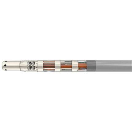
Defibrillating electrode: MRI compatible up to 3T, DF-4 connector, active fixation; presence of a steroid, standard electrode length of 59-64 cm,
maximum electrode diameter of 7.3 Fr.
Atrial electrode: MRI-compatible up to 3T, IS-1 Vi connector; active fixation, presence of a steroid, standard lengths of 45-59 cm, distance from the tip to the ring of no more than 11 mm, diameter of the electrode body of less than 2 mm.
Introducer discontinuous percutaneous, 2 pcs., sizes – 7, 8 Fr