


OPAL HDX MAP SYSTEM
Hybrid navigation technology based on magnetic and impedance information – simultaneous creation of an electromagnetic field map using a magnetic sensor and an electric field map containing impedance values that correspond to a coordinate in space.
Catheter localization accuracy in a magnetic field, (mm) is not worse than 1. Catheter localization accuracy for impedance tracking, (mm) is not worse than 2. The ability to use an ablation catheter from any manufacturer for application. The ability to build (or supplement) anatomy
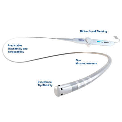
and/or electrical activity with an ablation or diagnostic catheter from any manufacturer, provided that there is a minimum anatomical volume built by a mapping (navigation) catheter. Compatibility with a high-resolution mapping (navigation) catheter that allows simultaneous recording of data from 64 electrodes to generate high-density electroanatomical maps. The ability to perform manual and automatic (continuous) mapping during sinus rhythm, tachycardia, or stimulation mapping. An intelligent algorithm for automatically annotating the mapped rhythm to create high-precision electroanatomical maps based on the inclusion/exclusion of specified criteria. Assessment of cycle length consistency. Assessment of interval consistency between two control points. Quantitative assessment of the morphology of the QRS complex in relation to a given template. Assessment of the constancy of respiratory dynamics – compensation for changes in the position of the catheter during the patient’s breathing in real time. Assessment of the constancy of the catheter’s movement during manipulation. Assessment of the stability of the catheter based on the comparison of the morphology of the electrogram signal with the previous one. Assessment of the quality of the localization of the magnetic and impedance system. Automatic selection of criteria for automatic annotation when selecting the targeted chamber and rhythm (sinus, tachycardia, or stimulation). The ability to include all points of the catheter’s position during anatomy mapping. The ability to include the catheter position point during the correct breathing phase. The ability to include the catheter position point when the specified criteria are met. The ability to create anatomical, isochronous (activation), isopotential (voltage), and fractionated maps. The ability to use isopotential (and isochronous) maps to accurately identify the area of post-infarction and/or post-surgical scarring. The ability to visualize the excitation wavefront on the anatomy of the heart chamber. The ability to map “areas of interest” in a single heart chamber. Function for measuring the distance between any points on the constructed map. Function for calculating the constructed volume.
Function of importing and integrating CT/MRI. Function of mapping based on anatomical maps created earlier. Program for viewing a three-dimensional model of the heart in any projection. Ability to create directional slices of the heart map at any angle and in any projection. Ability to record and view videos and screenshots taken during the procedure. Ability to mark major anatomical landmarks such as large vessels, valves, etc. Ability to save the electrograms of each point on the map for editing. Ability to create, edit, and store electroanatomical maps. The ability to record points of radiofrequency exposure. Simultaneous recording of both bipolar and unipolar signals. The ability to display the necessary EPHI channels on the screen. The ability to stimulate from any pair of electrodes on the reference catheter. The ability to stimulate from any pair of electrodes on the mapping catheter. The ability to display the parameters of radiofrequency exposure on the navigation system monitor for compatible RF generators. The ability to create maps of multiple heart chambers and display them simultaneously on the screen. The ability to adjust the degree of respiratory compensation during the procedure.
Display of the patient’s respiratory activity graph and the selected threshold of respiratory compensation on the navigation system monitor, with the ability to change it from this window during the operation. Ability to search for complex fractionalized atrial electrograms. Ability to hide and display points on the map. Local impedance calculation module for the ablation catheter. Module for displaying groups of signals on the map created by the high-density mapping catheter. Display of double potentials. Display of fractionalized potential areas. Displays the activation area during the selected cycle interval. Displays a histogram of the relative number of activation points over time within the mapping window. Displaying the local activation front on top of the isochronous map. An automatic ablation point annotation module based on specified ablation criteria. The criterion of stability of the catheter position. Criterion for changing the local impedance. Criterion for changing the impedance readings of a radio frequency generator. Displaying the ablation point in a different color depending on whether it meets or does not meet the specified criteria. The ability to display the distance to the nearest ablation point.
System components
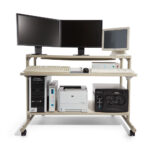
- OPAL / Rhythmia HDx™ Workstation (computer, keyboard, mouse)
- Linux-based operating system
- Signal station
- Dimensions: length x width x height, cm 41 x 38.9 x 26.2
- Input for 12 ECG leads
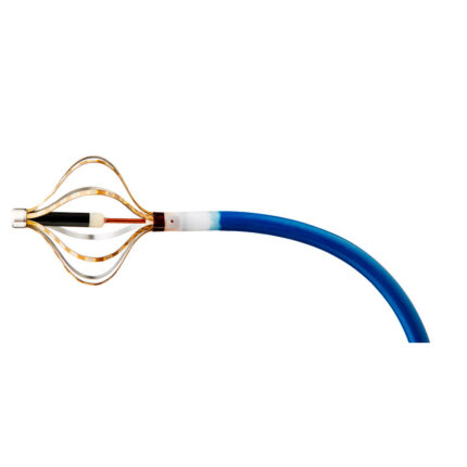
- Input for connecting a high-resolution mapping (navigation) catheter that allows simultaneous recording of data from 64 electrodes to produce high-density electroanatomical maps
- Connector for connecting a stimulator, channels 2
- Input for connecting the electrogram registration distribution unit 2
- Output – direct connection for transmitting signals from catheters to LSPro and GE Cardiolab electrophysiological systems
- Signal resolution, 24 bits
- Порог шума, мкВ 10
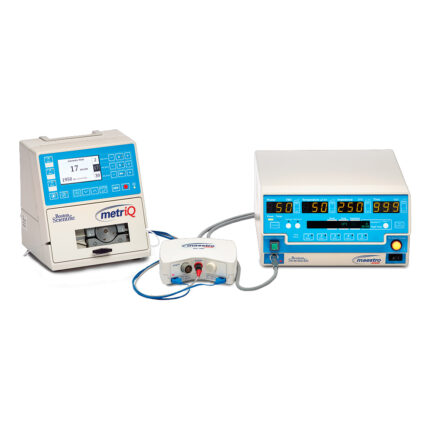
- Compatibility with modules for connecting to Stockert, Smartablate, and Maestro ablation generators
Magnetic navigation system
- Magnetic field emitter for localization of catheters with a magnetic sensor 1 piece
- 1 x Emitter Mount for the Operating Table
- Unit for connecting a navigated ablation catheter to the Maestro RF generator 1 piece
- Distribution unit for recording electrograms or output to the EFI system, 2 units
- 1 emitter connection cable
- 1 ECG signal acquisition cable
- 1 cable for connecting signals from the mapping system to the electrophysiological system
- 1 reference patch cable